
Causes and Risk Factors of Talar Dome Injuries
The talus is the ankle bone beneath the tibia (shinbone) and above the heel bone. Its rounded upper surface, the talar dome, is covered with cartilage that allows smooth movement at the ankle joint.
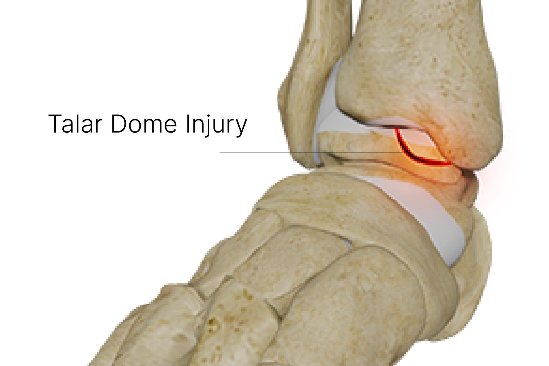
Ankle osteochondral lesions, particularly those in the talar dome, involve injuries to the cartilage and the bone beneath it. These lesions affect the upper joint surface where the talus meets the tibia and can cause pain, swelling or catching. The nature of talar dome injury symptoms and the necessary treatment vary, depending on whether the cartilage or the bone is affected, and can significantly influence daily life.
A talar dome injury affects the cartilage or underlying bone at the top of the talus, where it articulates with the tibia. Injury severity varies; symptoms and imaging guide management.
Talar dome injuries may follow an ankle sprain or other trauma, or develop with repetitive loading. Symptoms can persist after the initial injury. Foot shape or the surface someone trains on does not, by itself, establish the cause.
Persistent ankle pain, swelling or catching after an injury warrants assessment. Early evaluation can guide appropriate care, but recovery varies.
Tell Tale Signs & Symptoms of Talar Dome Injuries
Deep ankle pain during weight-bearing can occur with a talar dome injury, but the location of pain alone cannot distinguish it from a sprain or other ankle condition.
Swelling, stiffness, catching or locking may occur. Clicking alone is not diagnostic. A locked ankle or inability to bear weight needs prompt medical assessment.
Avoid activities that aggravate persistent ankle pain until it has been assessed. Treatment and return to sport depend on the lesion and your symptoms.
Podiatric Talar Dome Injury Management
Talar dome injuries can resemble other ankle problems. A podiatrist can assess symptoms and arrange medical or orthopaedic referral. X-rays and sometimes MRI or CT are needed to assess a suspected osteochondral lesion.
Early assessment can help identify the injury and guide treatment, although recovery and complications vary. Your podiatrist will help manage pain and symptoms and guide you in regaining strength and function in your foot. Don’t let a talar dome injury set you back. Seek professional advice and actively engage in your journey to optimal foot health.
Addresses Cartilage Damage for Osteochondral Lesions
For a stable osteochondral lesion, non-surgical care may include activity modification and a period of protected weight-bearing or immobilisation. This aims to reduce symptoms and loading; cartilage healing is not guaranteed.
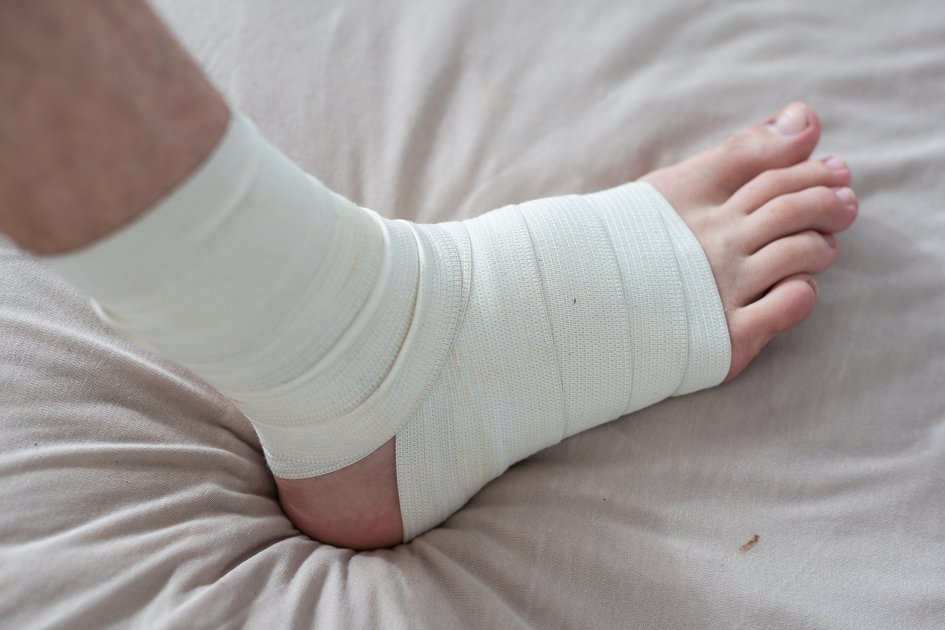
Ankle bracing can help stabilise and support the joint, maintaining alignment and reducing stress on the damaged area.
Specific physical therapy exercises that enhance ankle stability and strengthen the surrounding musculature are also key. This might include balance exercises, such as single-leg stands and proprioceptive training to enhance joint position sense.
Focus on Bone Injury for Subchondral Fractures
Subchondral fractures, on the other hand, are bone-related injuries. Conservative treatment here also starts with rest and immobilisation. Just like with cartilage injuries, rest is crucial. However, immobilisation might be more stringent, depending on the fracture severity.
A doctor or pharmacist can advise on pain relief and whether an anti-inflammatory medicine is suitable. These medicines may relieve symptoms but are not proven to restore damaged cartilage.
In both scenarios, it’s important to note that treatment effectiveness varies based on the injury’s specifics – the lesion’s size, exact location, and overall joint health. While these conservative treatments are often successful, particularly in early or mild cases, they require patience and adherence to the prescribed regimen.
More invasive approaches, including surgical intervention, may be considered when conservative management doesn’t yield the desired outcomes. The decision to escalate talar dome fracture treatment should always be made in consultation with a podiatrist and orthopaedic surgeon, ensuring a personalised approach based on individual needs and the injury’s characteristics.
Surgical Intervention for Talar Dome Injuries
Surgery may be considered for patients whose conditions are not alleviated by conservative methods or, in more severe cases, if their pain is debilitating and requires immediate treatment. Some surgeries are minor, like arthroscopy, where you can be discharged within the day. In this procedure, a surgeon uses an arthroscope to examine the joint from inside and potentially perform further surgical procedures.
Microfracture is one surgical option for selected lesions. Small holes in the underlying bone stimulate repair tissue, usually fibrocartilage, rather than restoring normal joint cartilage. Other procedures may be more appropriate for larger or unstable lesions.
Post-operative care follows the surgeon’s plan for weight-bearing, rehabilitation and return to activity. A podiatrist or physiotherapist may contribute to this coordinated care.
Schedule a Consultation to Manage Talar Dome Injury
Management of talar dome injuries is a delicate balance of allowing natural healing and actively encouraging recovery through targeted exercises and supports. By understanding the specific nature of the injury – whether it involves cartilage or bone – and following a structured treatment plan, most individuals can look forward to a return to function and activity. Remember, no matter how small, every step taken towards recovery is a move in the right direction.
If you suspect you have a talar dome injury, contact our podiatry centre today to make an appointment.