

Melanonychia is grey, brown or black pigmentation within a fingernail or toenail. It is caused by melanin, the pigment that gives colour to your skin and hair, inadvertently being deposited in the nail plate.
The most common pattern is called longitudinal melanonychia, which appears as a vertical band running from the base of the nail towards the tip. Many cases are harmless. However, a new or changing dark line, particularly on a single nail, needs assessment because nail melanoma (also called subungual melanoma) can cause melanonychia.
What Is Melanonychia?
Melanonychia describes general nail pigmentation rather than any one specific disease. It may affect a single nail, multiple nails or the whole nail plate. It can occur at any age and in any skin tone, though it is more common in people with darker skin tones.
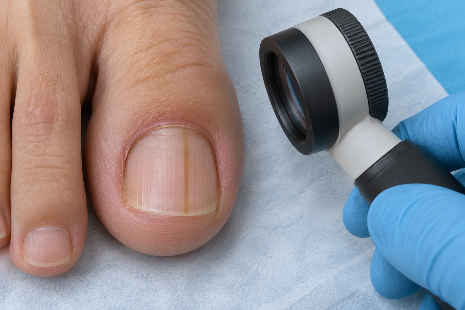
Melanonychia usually begins in the nail matrix, the tissue beneath the base of the nail that produces the nail plate. The nail bed supports the plate but is not its main growth centre. Two processes can cause melanonychia:
- Melanocytic activation: The number of melanocytes (pigment-producing cells) remains normal, but the cells themselves produce more pigment. Possible triggers include natural pigmentation, friction, trauma, nail biting, pregnancy, inflammation, infection and certain medicines.
- Melanocytic hyperplasia: The number of melanocytes increases. This may result from a benign mole or lentigo in the nail matrix. Less commonly, nail unit melanoma may cause it.
Melanonychia is not contagious. However, an infection that causes nail pigmentation may be contagious.
Types of Melanonychia
| Pattern | Appearance | Common context |
|---|---|---|
| Longitudinal melanonychia | A vertical brown, black or grey band extending from the nail base towards the tip | The most common pattern; also called linear melanonychia or melanonychia striata |
| Transverse melanonychia | A horizontal pigmented band extending across the nail | May be associated with medicines, inflammation or systemic factors |
| Diffuse or total melanonychia | Pigmentation involving most or all of the nail plate | May affect one or several nails, depending on the cause |
Longitudinal melanonychia may be narrow or broad and may involve one or more nails. A stable, regular pigmented band is often benign, particularly when similar bands affect multiple nails. Appearance alone cannot confirm the cause of melanonychia; an accurate diagnosis guides further management.
Symptoms
The main symptom is nail discolouration, often a dark line. The affected area may:
- Appear grey, brown or black.
- Form a vertical band (longitudinal melanonychia) or horizontal band (transverse melanonychia).
- Affect part of the nail or the entire nail plate.
- Involve one nail or several nails.
- Remain stable or gradually change in width, colour or shape.
Benign melanonychia is usually painless. Pain, bleeding, ulceration, splitting, nail distortion or a lump beneath or beside the nail requires medical assessment because another nail disorder may be present. These symptoms are very unlikely to be caused by melanonychia alone.
When to Seek Medical Care
Arrange an assessment for melanonychia if you notice any new or unexplained pigmented nail band. Seek care promptly if:
- A band is widening, darkening or changing rapidly.
- The dark band has uneven colour or irregular borders.
- Several shades of brown or black appear within the same band.
- Pigment extends onto the cuticle or surrounding skin.
- The nail becomes split, distorted, painful or prone to bleeding.
- A new dark line develops on one nail without a clear injury.
- The change affects the big toe, thumb or index finger.
- You have a personal or family history of melanoma.
- The pigmentation does not move towards the tip as the nail grows.
Pigment spreading from the nail onto the nearby skin is called Hutchinson’s sign. It can be associated with subungual melanoma, a form of skin cancer, but it does not confirm cancer by itself. Further assessment is necessary for an accurate diagnosis.
The ABCDEF guide
Healthcare professionals may use the ABCDEF guide when assessing longitudinal melanonychia for nail melanoma:
- A, age and background: The person’s age, skin type, medical history and risk factors.
- B, band: The brown-to-black band’s colour, width and border.
- C, change: Whether the band is widening, darkening or otherwise evolving.
- D, digit: Which finger or toe is involved.
- E, extension: Whether Hutchinson’s sign is present, with pigment extending onto the surrounding skin.
- F, family or personal history: A history of melanoma or atypical moles.
This guide helps identify concerning features, but it cannot diagnose or exclude subungual melanoma.
Causes of Melanonychia
Natural pigmentation
Melanonychia is more common in people with darker skin tones. Natural pigmentation often affects several nails and may remain stable for years. This is sometimes called racial melanonychia, also known as ethnic or constitutional melanonychia.
Friction or nail trauma
Repeated pressure from tight shoes, running or toe deformity can cause melanocytic activation in the nail matrix. Frictional melanonychia often affects toes exposed to repeated pressure.
Traumatic melanonychia is different from a fresh bruise between the nail plate and nail bed. A bruise contains trapped blood, while melanonychia contains melanin produced by the nail matrix.
Benign nail lesions
A mole or lentigo in the nail matrix can produce benign longitudinal melanonychia. A nail matrix nevus or mole is an important cause of melanonychia in children.
Medicines
Some chemotherapy medicines, antimalarial medicines and other systemic drugs can cause pigmented nail bands or diffuse nail darkening. Medication-related melanonychia may gradually fade after you stop the medicine, but you should never stop prescribed medicine without speaking to the prescriber.
Pregnancy and hormonal conditions
Hormonal changes during pregnancy can cause melanocytic activation, increasing pigment production. Endocrine disorders, including Addison’s disease, Cushing’s syndrome and some thyroid disorders, may also cause nail pigmentation, usually with other symptoms or pigmentation elsewhere on the body.
Inflammatory or systemic conditions
Psoriasis, lichen planus and other inflammatory nail disorders may cause melanocytic activation in the nail matrix. Vitamin B12 or folate deficiency, connective-tissue disease and some systemic illnesses have also been associated with melanonychia.
Infection
Fungal or bacterial nail infection may cause irregular brown pigmentation through melanocytic activation in the nail matrix. Fungal melanonychia may occur with thickening, crumbling, lifting or debris between the nail plate and nail bed. Testing may be needed because melanonychia alone does not confirm a fungal nail infection.
Nail unit melanoma
Nail melanoma is an uncommon but important potential cause of melanonychia. It may appear as a new band of longitudinal melanonychia on a single nail. The band may become wider, darker or more irregular. Pigment may extend onto the surrounding skin, and the nail may eventually split, lift, bleed or become distorted.
Most melanonychia is not melanoma. However, appearance alone cannot reliably exclude skin cancer.
Melanonychia or Another Nail Problem?
Black marks in a nail do not always contain melanin.
- Subungual haematoma: Blood becomes trapped between the nail plate and nail bed after an impact or repeated pressure. It may initially appear red, purple or black and usually moves towards the tip as the nail grows.
- Fungal nail infection: The nail may become yellow, brown, green or black, often with thickening, crumbling or debris.
- External staining: Nail polish, henna, dye, paint or chemicals may stain the nail plate.
- Thickened or damaged nail: Repeated pressure, psoriasis and previous nail injury may alter both nail colour and texture.
Read more about the broader causes of a black toenail.
Diagnosis
A healthcare professional will ask when melanonychia appeared, whether it has changed, how many nails are affected, and whether you have experienced nail trauma or repeated pressure from footwear. Your medical history may also include your medications, pregnancy, skin conditions, underlying medical conditions, and family history.
The examination includes the colour, width, borders, and distribution of melanonychia. The examiner also checks the affected nail plate, the proximal nail fold at the base, the lateral nail folds at the sides, the visible nail bed, and the surrounding skin.
Dermoscopy uses a magnifying instrument and specialised light to examine pigmented lesions and nail pigmentation. This may help distinguish a regular benign pigmented band from a pattern that needs further investigation, but dermoscopy alone cannot confirm nail melanoma.
If fungal infection is suspected, a nail clipping or scraping may be tested. If the findings are suspicious or remain unclear, you will be referred to a dermatologist or another doctor experienced in nail disease. A nail matrix biopsy provides tissue for histopathological examination and can establish whether nail melanoma or another pigmented nail lesion is present.
Management
Melanonychia management depends on the underlying cause.
Stable benign longitudinal melanonychia may only need clinical photography and periodic monitoring. Pressure-related melanonychia may improve when footwear fit, friction or repeated trauma is addressed. An infection or inflammatory disorder requires condition-specific care.
When a medicine may be responsible for melanonychia, the prescribing clinician can decide whether an alternative is appropriate. Do not stop a prescribed medicine without speaking to the prescriber.
Confirmed subungual melanoma requires specialist surgical and cancer care. Do not treat suspicious pigmentation cosmetically or cover it indefinitely without assessment.
Melanonychia in Children
Longitudinal melanonychia in children is usually benign and is commonly associated with a benign mole in the nail matrix or melanocytic activation. Features considered concerning for adults may appear in benign childhood lesions, so any assessment must be age-specific.
Depending on the pattern, a clinician may recommend photographic monitoring or specialist review. Biopsy is considered selectively because it can affect a child’s future nail growth.
Podiatry Assessment in Singapore
A podiatrist can examine a dark line on a toenail, document the pigmentation and assess possible footwear pressure, repeated trauma, fungal infection or another nail disorder. The assessment considers the number of affected nails, the pattern of longitudinal melanonychia, and the surrounding skin.
If the findings are suspicious or the cause of melanonychia remains uncertain, you may need a referral to a dermatologist or doctor for further investigation. A podiatrist cannot offer the diagnosis or assessments necessary when nail melanoma is suspected.
The Foot Practice assesses toenail changes as part of its skin and nail care services in Singapore. Call us today to arrange an assessment if a dark band is new, changing, unexplained or difficult to distinguish from a bruise or fungal infection.
Frequently Asked Questions
Should I be worried if I have melanonychia?
Not necessarily. Many cases of melanonychia are caused by natural pigmentation, friction, trauma or a benign nail lesion. However, a new or changing band, especially on a single nail, should be assessed because subungual melanoma and other pigmented nail disorders can look similar.
How do you get rid of melanonychia?
There is no single way to remove melanonychia because management depends on the cause. Benign melanonychia may not need treatment. Pigmentation caused by pressure, infection, medicine, or another underlying condition may improve when you address the trigger.
Can melanonychia fade away?
It may. Melanonychia related to trauma, infection, or certain medicines can gradually fade as a new nail grows. Natural brown pigmentation and benign nail lesions may persist. Toenails grow slowly, so visible improvement can take several months.
Can a B12 deficiency cause melanonychia?
Vitamin B12 deficiency is a reported cause of nail pigmentation, often affecting multiple nails and sometimes occurring with other symptoms. An isolated dark line does not confirm a deficiency. A healthcare professional can decide whether blood testing is appropriate.
Can melanonychia become melanoma?
Melanonychia describes nail pigmentation, not a growth that automatically becomes cancer. Most cases are benign. However, nail unit melanoma can present as melanonychia, which is why a new, widening, irregular or changing band needs assessment.